The technique of optical imaging measures changes in metabolism and blood flow from the brain surface. Instead of detecting changes in the electrical or magnetic structures of the neurons, like EEG, MEG and fMRI do, optical imaging observes changes in the reflectance of light from the brain surface.
Optical imaging is a technology that helps surgeons to map lesions in the body so that they can understand how certain conditions affect brain activity. This is an innovative tool for medical practitioners, that can shed extensive light on body lesions and the way how the brain operates.
How Optical Imaging Technology Works?
The technology uses light for extracting new information from the tissue, and inform and assist doctors to design and carry out surgeries for the removal of lesions. Basically, it is a kind of localization process in which this technology helps surgeons to pinpoint information about the location and depth of brain lesions precisely. This kind of information is not easily attainable with other technologies present currently.
The technology makes use of contrast for the absorption of light. Fluorescent agents are inserted into the body to find blood vessels and lesions within the tissues. The same kind of technology is also used for studying the activation of neurons in the brain, with the help of which doctors can detect conditions and diseases like Parkinson’s.
This technology overcomes a major challenge faced by fluorescence imaging, which is the scattering of light, due to which information received by the surgeon becomes limited. Optical imaging gives detailed information about the lesions for surgeons, which ultimately improves the results for the patients.
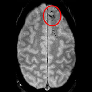
Identifying Brain Tumors and Lesions Using Optical Imaging Technology
With the help of optical coherence tomography imaging technology, malignant lesions in the brain can be diagnosed during surgery. This imaging technology takes images of the brain tissue, with the help of which surgeons can clearly differentiate between healthy and malignant cells. This information can be highly helpful in removing tumors in the future, thereby making them more efficient.
Brain lesions are one of the most common conditions related to the Central Nervous System. Sometimes, the lesion grows into the white substance of the CNS with no clear boundaries. In that case, OCT imaging technology can determine its boundaries, as it works on the principle of ultrasound diagnostics. Light wave penetrates into the tissue up to a couple of millimeters, and reflect it with intensity. This technology uses this data to create an image of the brain and the lesion.
The technology can diagnose multiple tissues, and it helps in identifying typical tissue characters to systematize them. As many as 300 images of the tissue can be taken during biopsy and tumor removal procedures of the patient. After that, they can classify them among healthy and malignant cells. However, the entire criterion depends on the intensity of the signals.
Light enters health brain tissues and dissipates them well. As a result, the surgeons receive intense signals from the white substance. Since tumor cells are fragmented, light passes through them farther and signals become less intense. As compared to MRI and ultrasound, OCT has high resolution and is more successful in identifying the homogeneity of the tissue structure. At present, researchers are working towards the improvement of its signal quality and making a special biopsy catheter.
Combination of Optical Imaging and Artificial Intelligence
Researchers are now combining the advanced method of optical imaging with Artificial Intelligence, with the help of which they will be able to produce a real-time, more accurate diagnosis of brain lesions. The technique reveals infiltration of tumor in the tissue by illuminating essential features that are not visible on standard histologic images, and it also collects scattered light. Then AI is used to process and analyze the microscopic images within minutes, and now surgeons can see predicted brain lesions diagnosis. With the help of the same technology, they can also detect accurately and remove an undetectable tumor.
With the help of this technology, surgeons can now see what would have been otherwise completely invisible. This improves the accuracy and speed of the OR and eliminates any risk of misdiagnosis. Some of the most common brain lesions and tumors include malignant glioma, metastatic tumor, meningioma, and lymphoma. The results derived from this technology are devoid of errors and are close to almost 100% accuracy. The system has a high diagnostic capacity, which is highly beneficial not only for the pathologists and surgeons but also for the patients.
We, at SepStream, have access to the best of imaging tools and technologies to deliver fast and accurate results. Whether you are looking to identify brain lesions, tumors, or anything else, our experts can help you during the process and give you the right guidance you would need to get the results right. So, get in touch with us today and know your options and alternatives.