Imagine, instead of a typical black and white X-ray picture, a physician reviews color images to identify the tissues being scanned! The color X-ray imaging technique is capable to produce more accurate and clearer pictures, assisting the doctors in giving a more precise diagnosis to their patients.
X-ray color images are coming into reality; thanks to MARS Bioimaging Ltd, a New Zealand-based company that, for the first time ever, scanned a human body using a revolutionary color medical scanner powered by Medipix3 technology developed at CERN. And the results are amazing! The breakthrough technology is capable of capturing high-quality 3D color X-ray images of the human body.
The new technology belongs to the family of Medipix, advanced read-out chips that are used for particle detection and imaging. It functions as a powerful camera, identifying and counting every particle that hits the pixels when the electronic shutter is open. This aids in generating high-resolution, reliable, and high-contrast pictures that make it a unique solution for various imaging applications, especially in the healthcare sector.
The new device incorporates hybrid pixel-detector technology that was developed initially for CERN’s Large Hadron Collider, which is “the world’s largest and most powerful particle accelerator. Medipix is the brainchild of father and son duo, who are scientists Professor Phil and Professor Anthony Butler from Canterbury and Otago Universities. They have spent decades in developing and refining the technology, building Medixpix3, which is the most advanced chip available today.
According to Professor Phil Butler, “this technology sets the machine apart diagnostically because its small pixels and accurate energy resolution mean that this new imaging tool is able to get images that no other imaging tool can achieve.” Mars Bioimaging Ltd is commercializing the next-generation 3D scanner and integrates the spectroscopic information produced by the Medipix3-enabled detector with critical algorithms to create three-dimensional color images.
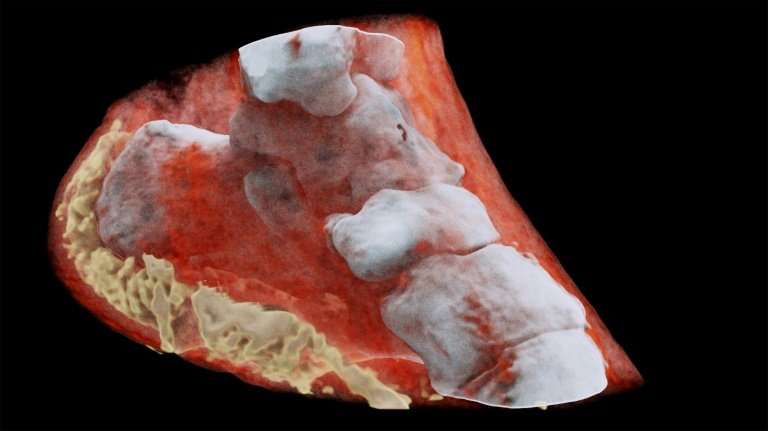
The detector, using its electronic shutter, identifies photos of various energy levels, whose energy signature allows the scanner to “differentiate and assign colors to a variety of tissues such as muscle, fat, water, and bones.” A computer algorithm then processes measurements of the photons to build 3D color images. Unlike grayscale X-ray pictures, full-color images will help physicians to perform a more accurate diagnosis. “Its small pixels and accurate energy resolution mean that this new imaging tool is able to get images that no other imaging tool can achieve,” said Professor Phil in a statement.
Till mid-July 2018, preliminary studies were conducted using a smaller version of the scanner to identify its results for cancer, joint health, bones, and vascular diseases that are responsible for strokes and heart attacks. “In all of these studies, promising early results suggest that when spectral imaging is routinely used in clinics it will enable more accurate diagnosis and personalization of treatment,” said Professor Anthony Butler.
In times to comes, researchers plan to scan rheumatology and orthopedic patients in New Zealand using the pioneering MARS scanner in a clinical trial that is the first ever in the world. This paves the way for high-quality radiology reporting, better diagnosis, and higher patient satisfaction.
SepStream® EMR/RIS/PACS Solutions further help streamline radiology workflow operations, offering a scalable and powerful cloud-based imaging system that meets your needs and reduces radiologist burnout.