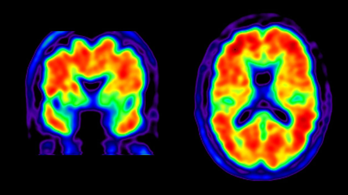
According to a study published on April 11, 2019, in the New England Journal of Medicine (NEJM), PET brain scans of former NFL players with neuropsychiatric and cognitive symptoms showed a high level of tau than controls in the brain regions that get typically affected by chronic traumatic encephalopathy (CTE).
The Background
Chronic traumatic encephalopathy (CTE) is a typical neurodegenerative disease associated with a history of repeated head impacts. The process of neuropathological diagnosis is primarily based on specific patterns of tau deposition and minimal amyloid-beta deposition, which differs from other disorders such as Alzheimer’s disease. However, the feasibility to detect tau and amyloid deposition within the living brains of human beings at risk of CTE is yet to be studied.
The study used flotaucipir positron-emission tomography (PET) and florbetapir PET for measuring the deposition of tau as well as amyloid-beta in the brains of former NFL (National Football League) players having cognitive disabilities and neuropsychiatric symptoms as well as in asymptomatic men with no relevant signs of traumatic brain injury or accident.
Automated image analysis based algorithms have been used for comparing the local standardized uptake value ration, or SUVR – the ratio of radioactivity in the cerebral region to that present in the cerebellum) between these two groups and further explore the relationship between SUVR and severe symptoms and years of playing football in the former group of players.
The Result
Flortaucipir PET showed high amounts of tau present in three parts of the brain – bilateral superior frontal region, left the parietal region, and bilateral medial temporal region of 26 former players of the NFL compared to the 31 healthy controls without any history of traumatic injury of the brain.
Robert A. Stern Ph.D. Boston University School of Medicine and colleagues wrote, “The group of living former NFL players with cognitive, mood and behavioral symptoms who were examined in this study had elevated flortaucipir PET measurements in regions of the brain that were consistent with the distribution of tau in persons with CTE confirmed by postmortem examination.”
The authors also added, “Detection of the disease during life could be used to assess its epidemiology, risk factors, and course and could be used in treatment and prevention trials.”
Typically, these types of tau PET changes related to CTE are only measurable at the time of postmortem exams. This new finding could influence or change the approach of clinicians towards this disease.
In addition, the authors also performed an exploratory analysis to find correlation coefficients between 0.45 and 0.58 for the total number of years of playing football and the tau deposition level.
According to reports, tackle football has dramatic effects on the development of the brain of young players. Some neuropathological studies involving former football players found that the number of years of playing tackle football is associated with the nature and severity of tau deposition.
Tackle Football Study
Just one season of tackle football may cause changes in the development of the brain of younger players, reports an RSNA study in Chicago in 2018.
The study included 670 youth and high school football players without a history of developmental, psychiatric or neurological abnormalities, and no concussions before, during or after the tackle football session. To determine is repeated head injury affected the normal pruning of the brain; each of those 60 players was given a Head Impact Telemetry System (HITS) support helmet. The customized helmet was lined with sensors called accelerometers, which could measure the impact data and detect the concussion risk of a player.
Final Procedure
The complete research did not reveal a high proportion of positive florbetapir PET scans or even higher amyloid-beta deposition in comparison to controls.
The authors of the study wrote, “These findings suggest that the cognitive difficulties reported by the former players were not related to Alzheimer’s disease amyloid-beta deposition.
Allan H. Ropper, MD, Deputy Director NEJM also stated in an editorial that like Alzheimer’s disease, the science behind CTE is “in a phase of fumbling with circumstantial evidence for a connection between tau deposition and clinical syndrome.”
He also added, “The report in this issue certainly does strengthen the case that tau is the offender early in CTE, but other links remain to be clarified. The techniques for studying living biology, such as this use of tau-ligand PET, are making a difference.
Avid Radiopharmaceuticals funded this study partially.
sepStream® uses only advanced and highly efficient imaging tools for an accurate diagnosis. As imaging like CT scan, PET scan and MRI are critical instruments for detecting major complications; they only use the latest technology and equipment to deliver positive results at a much faster pace. It is the utmost endeavor of the team to provide sophisticated diagnostic tools and imaging results for improved healthcare. With faster turnaround time and accuracy, it may be possible to rule out major risk factors or gaps in reporting.