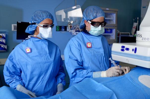
X-ray is considered an essential tool to conduct medical examinations of the patients undergoing diagnostic radiology. Most diagnostic examinations are based on effective X-ray system, which is acknowledged crucial for enhancing human health. However, it also poses the highest levels of health risks caused by radiation exposure. Radiation safety of the patients is also breached during Positron Emission Tomography when the body is exposed to high levels of “gamma radiation energy of positron-emitting isotopes.”
During the last decade, Digital Radiographic technology has been harnessed to minimize the potential risks of radiation exposure, while improving image utility. However, it fails to address the situations of unwanted radiation exposure caused due to the operator’s lack of understanding or inexperience. Therefore, initiatives are continuously being taken to adopt optimal radiation practices and ensure effective monitoring to reduce radiation dosage.

Radiation Monitoring Technologies Can Help Reduce Dose
Effective monitoring of radiation dosage and medical imaging practices is crucial to reduce patient and operator exposure. Several radiation monitoring technologies are available nowadays that use Digital Imaging and Communications in Medicine (DICOM) data to set up a structured dose report. “This system utilizes the radiation dose data-archiving method of standard DICOM dose SR combined with a DICOM modality performed procedure step (MPPS).”
“Digital radiographic systems contribute to patient radiation exposure information archives, thus also supporting digital information and communications in medicine (DICOM) header information, modality performed procedure step (MPPS) technology, and DICOM dose structured reports,” researchers suggest. The Integrated Healthcare Enterprise has published a document on radiation dosage monitoring to initiate the DICOM standards. The document also highlighted the importance of having a national radiation dose registry because the same radiology examination leads to 10-30 times fluctuations in the amount of radiation exposure when performed at different institutions.
Then there are systems that aid in “non-fluoroscopic localization of the catheters and the creation of 3D images during Electrophysiology (EP) procedures. Magnetic Resonance Imaging can be harnessed offline to produce high-quality 3D images without causing radiation overdose. Even in X-rays, the use of modern technologies such as pulsed fluoroscopy, filtration, real-time digital fluoro-processing, collimation, etc. can help minimize radiation dose dramatically.
However, technology alone cannot minimize radiation exposure of the patients in clinical procedures. It is vital for physicians to know how such technology can be leveraged for effective monitoring of radiation dosage. In a recent study, the researchers have analyzed the level of radiation exposure at different working locations. Based on their study, they have suggested optimal radiation levels for imaging operators and highlighted the importance of having “good radiation practices.”
According to the study, “Use of radiation protection devices such as L-bench reduces exposure significantly. PET/CT staff members must use their personnel monitors diligently and should do so in a consistent manner so that comparisons of their doses are meaningful from one monitoring period to the next.” Similarly, effective radiation monitoring practices need to be undertaken to reduce patients’ dose.
sepStream® offers affordable cloud-based EMR/RIS/PACS solutions that help digitize your imaging workflow, reduce radiologist burnout, improve patient satisfaction, and ensure cost-savings.