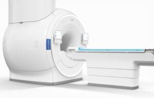
The U.S. Food and Drug Administration, more commonly known as the FDA, has given Philips 510(k) complete clearance for its much talked about MRI platformwhich is powered by Artificial Intelligence and is designed for the treatment of cancer that has been developed in one’s neck or head.
This particular software gives clinicians the opportunity to polish radiotherapy planning pertaining to the soft tissue tumors of the neck and head. The software also discards the requirement for imaging exams which include doing CT scans. This used to be done by extracting CT-like images from a standalone MRI scan that takes just about three minutes to be completed.
Ilya Gipp, who serves as the chief medical officer in charge of oncology solutions at Philips, has shared some details about the clearance that was given to them recently. According to Ilya, MR’s soft tissue imaging, along with the integration and orchestration of information with the help of artificial intelligence, will result in better clarity in radiotherapy planning.
MRCAT, enabled by AI, features important applications pertaining to the effective treatment of brain cancer along with cancer developed in the prostate and pelvis. It also has the potential to make processes like patient positioning and dose calculations easier.
This particular clearance was the subject of discussion at the recently organized annual meeting for the members of the American Society for Radiation Oncology. During this meeting, Philips confirmed that the organization has joined hands with MacroMedics, a reputed company that is known for its work in the space of patient positioning solutions put together for radiotherapy treatments.
In an official press release, Gipp stated that Philips has collaborated with MacroMedicsto create a mask system that would be patient-friendly and compatible with the company’s dStreamimaging coils. This, according to Gipp, is a part of Philips’ endeavor to offer the kind of precision tools required for the characterization and localization of tumors that are considered to be very difficult to treat.
What You Should Know About Radiotherapy Planning
Radiation Treatment Planning or RTP refers to the process which involves an elaborate team consisting of medical physicists, radiation therapists, radiation oncologists and medical dosimetrists making plans for the internal brachytherapy treatment procedure or external beam radiotherapy that a cancer patient needs to undergo.
When Did It Start?
When radiotherapy was first discovered, it was carried out on 2D X-Ray images with the help of manual calculations done by a physicist or an oncologist. In the 1970s, computerized treatment planning procedures started being used to enhance the overall speed and accuracy of dose calculations.
As one stepped into the ‘90s, powerful computer systems announced their arrival. CT scans were discovered and dose calculation algorithms also witnessed massive improvement. Because of all this, radiotherapy planning got better.
3D conformal planning or 3DCRT was also discovered around the same time and listed as a Level 2 procedure European Dynarad consortium. MLCs are used by 3DCRT to design the radiotherapy beam in a way that resembles the target tumor’s shape. This results in the healthy surrounding tissue’s dose getting reduced.
VMAT and IMRT, which are techniques belonging to level 3, carry out inverse planning to enable dose distributions in an improved manner. These types of methods are getting more popular, especially when it comes to the treatment of cancers.
What is Image Guided Planning?
Medical imaging is used extensively to put together a virtual patient for a design-based procedure that will be carried out with the help of a computer system. As far as treatment planning is concerned, a CT scan is chosen to be the primary image. Magnetic resonance imaging, on the other hand, offers a secondary image designed for the purpose of soft tissue contouring.
Positron emission tomography is not used very frequently and is largely set aside for cases where certain uptake studies can enable the process of making plans for target volume delineation. Contemporary treatment planning systems offer you tools that prove to be effective in multimodality image matching which is also referred to as image co-registration.
Treatment simulations prove to be helpful in planning the radiological, dosimetric and geometric aspects pertaining to the therapy with the help of radiation transport simulations. When it comes to intensity-modulated radiation therapy or IMRT, this particular procedure involves choosing a beam-type energy and making other physical arrangements.
- Forward Planning
When it comes to forward planning, beams are placed into a radiotherapy planning system by the planner. This system is designed to provide radiation to a tumor while making sure the healthy tissue receives a minimal dose.
Here, one has to take certain important decisions including the number of radiations beams to be given, the angles they will be delivered and whether attenuating wedges are required to be used or not.
- Inverse Planning
Inverse planning involves a radiation oncologist defining the tumor and critical organs of a patient. After this, the planner offers target doses and lays out the pertinent factors related to the procedure. An optimization program is carried out to arrive at the treatment plan that would complement the input criteria.
If you are looking for high-quality software solutions that are offered at an affordable price point, you should get in touch with sepStream®. They also provide imaging upgrades for healthcare facilities and clinics.