A lot of individuals suffer from Lyme disease. It is one of the most widespread vector-borne infectious diseases. 10-20% of people deal with post-treatment Lyme disease symptoms in spite of the necessary antibiotic treatment. Cognitive issues are evident among patients with PTLD, indicating that the disease, when it persists for a certain period, can impact and cause changes in the brain.
According to the latest research, individuals who suffer from Lyme disease for a considerable number of days develop functional and structural brain abnormalities. Both are closely associated with cognition and can make patients experience cognitive difficulties.
John Hopkins Medicine Researchers’ Report
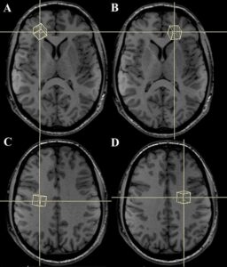
Credible researchers who are with John Hopkins medicine used the functional Magnetic Resonance Imaging technique. They did so, along with making patients perform specific tasks pertinent to short-term memory. The sole objective of doing so is to visualize the activity of the brain in those who had already contracted and suffered from Lyme disease and underwent treatment.
The researchers noticed and mentioned in the report that there were noticeable changes, primarily in the brain’s white matter. Even they found issues in post-treatment Lyme disease-affected people’s brain tissue physiology.
Post conducting the research, the team of notable researchers confirmed that MRI scans validate that people with PLTD experience cognitive and memory-related issues. And these health issues are associated with structural and functional changes in their brain.
Steps the Researchers Had to Take for the Study
The team of researchers took 12 people into consideration who were affected by PTLD and 18 individuals who were not initially diagnosed with Lyme disease. However, both groups were undergoing functional MRI exams.
Right after conducting the specialized imaging tests, they could find abnormal activities, mainly in the brain’s frontal lobe. This particular part of the brain helps humans perform cognitive tasks like concentrating on things or recalling a memory.
The researchers asked participants from each group to recall after memorizing lowercase and uppercase letters besides multiple letters’ alphabetical order. The imaging findings confirmed that there were distinctive differences between both groups’ frontal lobe activities in the brain’s white matter. The issue was found operating with relatively less flow of blood than gray matter.
White matter plays an instrumental role in transmitting information all around the brain. It acts as a rail track, assisting in disseminating information to the gray matter. The researchers conducted diffusion tensor imaging tests on every participant. They noticed that cognitive function was better in patients who already underwent treatment for Lyme disease.
Nowadays, hospitals and diagnostic centers in considerable numbers use SepStream® software to conduct imaging exams on patients to obtain accurate reports. Errorless imaging results help radiologists interpret MRI scans without confusion and keep patients updated on their conditions.