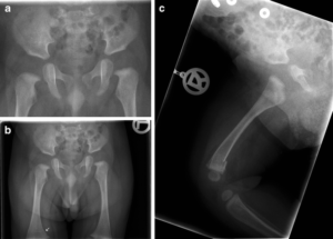
Imaging has always been one of the most critical requirements and tools of the diagnostic medical care lateral. When the radiologist and the physicians get to see the picture clearly, understanding the disease, its stage, and pace of advancement become easier and more accurate.
Therefore, the treatment process also improves and the patient experienced an augmented level of medical care. Precision in imaging is critical for all diseases, fatal or less damaging. Like many other domains of the medical care sector, imaging, too, is going through a digital up-gradation.
Collimating is a well-known process in the field of imaging the experts use before exposing the patient’s body to radiation for imaging.
Collimating anatomy enhances the image quality by multiple times. However, with digitized methods gradually taking hold of the imaging and radiology processes, collimating has also undergone a technological change.
The new method introduced involves digital cropping of the images following their exposures. The entire process takes place in the workstation.
Even though digital cropping appears to be a harmless process without any noticeable consequences on the outcomes, a team of researchers associated with the latest study has opined otherwise. The enlightening piece of work got published in the Journal of Medical Radiation Sciences recently.
It has explained the impact of digital cropping in detail claiming that the process is not without its impacts.
Sally Ball works with Australia’s Princess Alexandra Hospital and was one of the authors of this recent study. According to this new paper, published by Sally and her colleagues, digital images, lacking collimation, often turns out to be of inferior quality.
Therefore, a higher number of scatter hits the digital detector. The team further suggested that as the number of scatters increases, the histogram widens invariably. As a result, the image turns greyer and the spatial resolution diminishes.
Finally, due to all these impacts, the image loses a significant part of detailing pertaining to the anatomy concerned.

The Study
The experts initially focused on comprehending how frequently or extensively radiographers can use electronic cropping. They also conducted a study to understand how such a practice can change the quality of the image.
To carry out the study, the team of researchers chose 359 patients and took their musculoskeletal radiographs. The team compared the images taken before the cropping with the cropped versions. They were trying to figure out if the exposed area was bigger than a certain reference benchmark.
The Observation
The researchers had five projection points – AP shoulder, AP knee, lateral cervical spine, horizontal beam (lateral hip), and lateral facial bones. They dealt with 1071 measurements in this process. The data revealed that 61.2% of measurements exceeded the standard reference level.
This implied that the images went through electronic cropping and were not collimated before image acquisition.
Final Suggestion
No matter the impact, the researchers were not in favor of discarding the entire process of cropping keeping the trend of digitization in mind. The experts believe that sooner or later, digitization will take over the medical processes existing today.
Additionally, the team also focused on cropping’s time-saving aspect. They also mentioned that the scope of improvement of this process seems brighter through more innovations and digital audits.
Therefore, more research and efforts can develop more efficient cropping methods without jeopardizing the image quality the least.
At SepStream®, we understand how critical every image remains for the patients and the physicians attending them. From detecting the disease properly to offering the best treatment course, everything relies on these images.
We offer a variety of AI-integrated software solutions for accurate, fast and crystal-clear imaging. With our software solutions, providing premium-grade medical care to all patients become effortless.