A recent study has revealed the significance of contrast-enhanced mammography as an alternative to neoadjuvant chemotherapy. Especially for breast cancer patients, breast MRI has always been one of the methods for diagnosis. A recent study has suggested contrast-enhanced mammography as a better option after assessing the response of cancer patients.
The Study
The study also revealed that MRI and contrast-enhanced mammography provided for comparative evaluation of the size of the lesion. The researchers conducted the study on a sample of 51 patients. However, both the methods had equivalent specificities for pCR (pathologic complete response).
Both the methodologies of MRI and contrast-enhanced mammography gave an overestimation of lesion size. On the other hand, pathology provided a better measure of the lesion size. However, experts further clarified that both methods CEM and MRI can be critical for monitoring the treatment responses at different levels.
The Outcomes & Analysis
Rubina Manuela Trimboli works with Milan’s IRCCS Humanitas Research Hospital. She co-authored the study and mentioned that MRI is more sensitive to pCR. She further explained that a delay in CEM acquisition may aid in the process of residual DCIS detection.
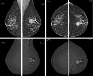
The study involved 51 patients with a mean age of 46 years. They all had biopsy-backed breast cancer and qualified for NAT or neoadjuvant therapy. These women had the experience of MRI and CEM before, during, and after their courses of treatment. The CEM, done after the treatment, had a delayed acquisition by six minutes.
Two experienced radiologists analyzed the outcomes. One of them had extensive experience in analyzing breast MRIs and the other CEM. They considered the outcomes carefully for the presence and size of the growing lesion. They also looked for the absence or presence of DCIS or invasive cancer.
Post-treatment pCR analysis of CEM gave 83% and 81% sensitivity. The percentage figures changed to 81% and 89% in the case of a delayed acquisition. The sensitivity and specificity of post-treatment MRI turned out to be 100% and 86%. MRI, CEM, and delayed CEM overestimated the size of residual tumor during post-treatment imaging by 1.9mm, 0.8mm, and 1.2 mm, respectively.
Since the results were pretty comparable, the experts suggested CEM when MRI examination is not possible.
MRI has always remained a reliable choice for NAT monitoring. However, researchers consider CEM a fitting alternative when MRI is not feasible. They concluded so based on the recent study.
Imaging solutions often offer the easiest ways to treat diseases like cancer. At sepStream®, we offer advanced imaging software solutions based on intuitive technology. Our AI-integrated solution ensures zero delay and zero flaw diagnosis. We remain committed to offering only the best quality solutions at affordable rates.