Parkinson’s disease is a neurological condition with no categorical treatment or sure-cure ascribed to it as of yet. While curing the condition is not possible as of now, restricting its symptoms and advancement is possible. Especially if the medical diagnostic system can ensure early detection of the disease, many patients can benefit enormously.
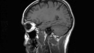
Quantitative magnetic resonance is precisely the way proposed by the researchers for the same. The researchers recently found out that quantitative MRI can trace the initial structural changes in the neurological system, indicating a future onset of the disease.
The researchers developed an analysis method for coming to this conclusion. They focused on striatum imaging in their study.
When Parkinson’s sets in, this part of the brain generally deteriorate. Therefore, the waning of the striatum has a direct association with the appearance of Parkinson’s.
The Study
Prof. Aviv Mezer, the lead author of the study, works in Jerusalem’s Hebrew University’s Edmond and Lily Safra Center of Brain Sciences. He and his colleagues explained in their article that a holistic understanding of basal ganglia functionalities is possible by mapping the spatial changes taking place in the striatum.
The team of researchers chose 23 participants for the study from an age group of 23 to 31. The sample also included 18 more participants aged between 55 and 75. Each of these participants went through quantitative MRI before an elaborate, multi-layer imaging and cognitive analysis.
The Outcome
The detailed imaging analysis unearthed unusual putamen gradients in individuals suffering from Parkinson’s. The gradients were particularly noteworthy for posterior putamen. The researchers found this result critical for explaining conditions like motor dysfunction and dopaminergic loss in Parkinson’s patients.
The team of researchers further explained that in their attempt to generalize the clinical MRI analysis, they found the in-vivo micro-structural correlates of PD. The team associated this correlate with both motor function abnormalities and dopamine loss faced by Parkinson’s patients. The team further found explicit reliance between cortical hierarchy and striatal position and explained the cortico-striatal relationships thereby.
Since the study had young and older participants, the team of researchers could compare the striatum’s micro-structural changes based on ageing. The team found that normal aging has a connection with asymmetry amplifications in the caudate’s posterior and anterior segments. On the other hand, patients with Parkinson’s disease showed putamen’s posteriors segment alterations.
The team of researchers found this series of revelations quite stimulating. They plan to implement this method to analyze the microstructure of several other parts of the human brain. The researchers are also finding novel ways to improve qMRI to use it for clinical practices. The team hopes to complete this work in another five years at most.
At sepStream®, we offer affordable imaging solutions keeping the modern requirements of the medical care domain in mind. Our AI-integrated intuitive imaging software ensures fast and flawless detection of diseases. Early and accurate detection enhances the quality of medical care provided by physicians and clinics. Go through our gamut of software solutions to choose the fittest one for you.