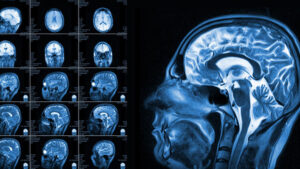
According to a new research study by a reputed neuroscientist from an esteemed University in Chicago, individuals who commit murders have reduced gray matter. Mainly in the brain’s regions linked with social cognition, behavioral regulation and emotional processing, reductions in gray matter were evident. The same condition was not apparent in people guilty of other offenses.
The study also emphasizes that the way a murderer processes morality or empathy varies notably from the ones that commit other crimes. As per the neuroscientist, if there were more gray matter, meaning more neurons, cells and glia, a homicide could better control his or her behavior. Ultimately, they could process emotional information better and restrain themselves from reacting impulsively.
The Jury is yet to give a Final Verdict on Parker Case
Taylor Parker was found guilty of capital murder on 3rd October by a body of judges in Texas. She was put behind bars for committing criminal offenses in the month of October 2020. The jury is yet to make their final decision on whether, because of the homicide, Parker will remain imprisoned until death.
The situation is so at present because Parker’s defense attorney brought a neuropsychologist to the stand during her trial at court. The healthcare professional conducted several neuroimaging examinations on Parker and found chronic dysfunction in her brain.
As per the medical expert’s interpretation of the neuroimaging findings, the convict has lost the ability to control her reaction tendencies because of severe dysfunction. The brakes of inhibition become numb when a person’s frontal and temporal lobes stop functioning properly.

Another Medical Professional’s Review of Parker’s Imaging Findings
Besides evaluating medical records and neuroimaging findings thoroughly, Dr. Siddartha Nadkarni conducted an assessment on his own to understand Parker’s condition better. He performed a series of brain scans along with an EEG. Inferences he has drawn after performing MRI, and other necessary imaging tests confirm that the convict suffers major dysfunction of frontal and temporal lobes.
According to different Texas news portals, Dr. Nadkarni mentioned that individuals having frontal lobe dysfunction are unable to apply the brakes of inhibition to keep themselves in control. They are impulsive and react promptly without any cognitive reasoning.
In addition, the healthcare expert said that he found damage in multiple parts of the convict’s brain. He pointed out the drying up of the white matter in the parietal lobe of Parker, besides significant problems in the frontal cortex.
All these severe issues in her brain have had an adverse impact on her behavior. The medical professional has mentioned that because of this sort of tissue wastage, such people suffer frequent irritability, apart from experiencing problems related to inhibition, memory and consistency.
Prosecutors’ Take after Reviewing Defendant’s Neuroimaging
In order to plead the case for their own sake, prosecutors felt the urge to use the neuroimaging findings of the defendant. According to prosecutors, they consulted certified physicians who found no severe issues in Parker’s brain and considered the scans quite normal.
The neuro specialist pointed out that opinions did vary because of different interceptions of the homicide’s brain scans. He could find several indicators that prove brain dysfunction. On the other hand, the doctors with whom the prosecutors got in touch looked for stroke-related indications.
The majority of diagnostic centers rely on sepStream® solutions these days. The prime reason is this software helps eliminate confusion by furnishing highly accurate neuroimaging results or reports quickly. Doctors and patients’ near and dear ones can have easy access to the imaging report no matter where they are, and everyone can intercept it in the correct manner.